Human body
| Human body | |
|---|---|
 | |
| Details | |
| Identifiers | |
| Latin | corpus humanum |
| MeSH | D018594 |
| TA | A01.0.00.000 |
| FMA | 20394 |
| Anatomical terminology | |
The human body is the structure of a human being. It is composed of many different types of cells that together create tissues and subsequently organ systems. They ensure homeostasis and the viability of the human body.
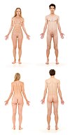
It comprises a head, neck, trunk (which includes the thorax and abdomen), arms and hands, legs and feet.
The study of the human body involves anatomy, physiology, histology and embryology. The body varies anatomically in known ways. Physiology focuses on the systems and organs of the human body and their functions. Many systems and mechanisms interact in order to maintain homeostasis, with safe levels of substances such as sugar and oxygen in the blood.
The body is studied by health professionals, physiologists, anatomists, and by artists to assist them in their work.
Composition[edit]

The human body is composed of elements including hydrogen, oxygen, carbon, calcium and phosphorus.[1] These elements reside in trillions of cells and non-cellular components of the body.
The adult male body is about 60% water for a total water content of some 42 litres. This is made up of about 19 litres of extracellular fluid including about 3.2 litres of blood plasma and about 8.4 litres of interstitial fluid, and about 23 litres of fluid inside cells.[2] The content, acidity and composition of the water inside and outside cells is carefully maintained. The main electrolytes in body water outside cells are sodium and chloride, whereas within cells it is potassium and other phosphates.[3]
Cells[edit]
The body contains trillions of cells, the fundamental unit of life.[4] At maturity, there are roughly 30[5]–37[6] trillion cells in the body, an estimate arrived at by totaling the cell numbers of all the organs of the body and cell types. The body is also host to about the same number of non-human cells[5] as well as multicellular organisms which reside in the gastrointestinal tract and on the skin.[7] Not all parts of the body are made from cells. Cells sit in an extracellular matrix that consists of proteins such as collagen, surrounded by extracellular fluids. Of the 70 kg weight of an average human body, nearly 25 kg is non-human cells or non-cellular material such as bone and connective tissue.[5]
Cells in the body function because of DNA. DNA sits within the nucleus of a cell. Here, parts of DNA are copied and sent to the body of the cell via RNA.[8] The RNA is then used to create proteins which form the basis for cells, their activity, and their products. Proteins dictate cell function and gene expression, a cell is able to self-regulate by the amount of proteins produced.[9] However, not all cells have DNA – some cells such as mature red blood cells lose their nucleus as they mature.
Tissues[edit]
 | |
The body consists of many different types of tissue, defined as cells that act with a specialised function.[10] The study of tissues is called histology and often occurs with a microscope. The body consists of four main types of tissues – lining cells (epithelia), connective tissue, nervous tissue and muscle tissue.[11]
Cells that lie on surfaces exposed to the outside world or gastrointestinal tract (epithelia) or internal cavities (endothelium) come in numerous shapes and forms – from single layers of flat cells, to cells with small beating hair-like cilia in the lungs, to column-like cells that line the stomach. Endothelial cells are cells that line internal cavities including blood vessels and glands. Lining cells regulate what can and can't pass through them, protect internal structures, and function as sensory surfaces.[11]
Organs[edit]
Organs, structured collections of cells with a specific function,[12] sit within the body. Examples include the heart, lungs and liver. Many organs reside within cavities within the body. These cavities include the abdomen and pleura.
Systems[edit]
Circulatory system[edit]
The circulatory system comprises the heart and blood vessels (arteries, veins and capillaries). The heart propels the circulation of the blood, which serves as a "transportation system" to transfer oxygen, fuel, nutrients, waste products, immune cells and signalling molecules (i.e., hormones) from one part of the body to another. The blood consists of fluid that carries cells in the circulation, including some that move from tissue to blood vessels and back, as well as the spleen and bone marrow.[13][14][15]
Digestive system[edit]
The digestive system consists of the mouth including the tongue and teeth, esophagus, stomach, (gastrointestinal tract, small and large intestines, and rectum), as well as the liver, pancreas, gallbladder, and salivary glands. It converts food into small, nutritional, non-toxic molecules for distribution and absorption into the body.[16]
Endocrine system[edit]
The endocrine system consists of the principal endocrine glands: the pituitary, thyroid, adrenals, pancreas, parathyroids, and gonads, but nearly all organs and tissues produce specific endocrine hormones as well. The endocrine hormones serve as signals from one body system to another regarding an enormous array of conditions, and resulting in variety of changes of function.[17]
Immune system[edit]
The immune system consists of the white blood cells, the thymus, lymph nodes and lymph channels, which are also part of the lymphatic system. The immune system provides a mechanism for the body to distinguish its own cells and tissues from outside cells and substances and to neutralize or destroy the latter by using specialized proteins such as antibodies, cytokines, and toll-like receptors, among many others.[18]
Integumentary system[edit]
The integumentary system consists of the covering of the body (the skin), including hair and nails as well as other functionally important structures such as the sweat glands and sebaceous glands. The skin provides containment, structure, and protection for other organs, and serves as a major sensory interface with the outside world.[19][20]
Lymphatic system[edit]
The lymphatic system extracts, transports and metabolizes lymph, the fluid found in between cells. The lymphatic system is similar to the circulatory system in terms of both its structure and its most basic function, to carry a body fluid.[21]
Musculoskeletal system[edit]
The musculoskeletal system consists of the human skeleton (which includes bones, ligaments, tendons, and cartilage) and attached muscles. It gives the body basic structure and the ability for movement. In addition to their structural role, the larger bones in the body contain bone marrow, the site of production of blood cells. Also, all bones are major storage sites for calcium and phosphate. This system can be split up into the muscular system and the skeletal system.[22]
Nervous system[edit]
The nervous system consists of the central nervous system (the brain and spinal cord) and the peripheral nervous system consists of the nerves and ganglia outside the brain and spinal cord. The brain is the organ of thought, emotion, memory, and sensory processing, and serves many aspects of communication and controls various systems and functions. The special senses consist of vision, hearing, taste, and smell. The eyes, ears, tongue, and nose gather information about the body's environment.[23]
Reproductive system[edit]
The reproductive system consists of the gonads and the internal and external sex organs. The reproductive system produces gametes in each sex, a mechanism for their combination, and in the female a nurturing environment for the first 9 months of development of the infant.[24]
Respiratory system[edit]
The respiratory system consists of the nose, nasopharynx, trachea, and lungs. It brings oxygen from the air and excretes carbon dioxide and water back into the air.[25]
Urinary system[edit]
The urinary system consists of the kidneys, ureters, bladder, and urethra. It removes toxic materials from the blood to produce urine, which carries a variety of waste molecules and excess ions and water out of the body.[26]
Anatomy[edit]
Human anatomy is the study of the shape and form of the human body. The human body has four limbs (two arms and two legs), a head and a neck which connect to the torso. The body's shape is determined by a strong skeleton made of bone and cartilage, surrounded by fat, muscle, connective tissue, organs, and other structures. The spine at the back of the skeleton contains the flexible vertebral column which surrounds the spinal cord, which is a collection of nerve fibres connecting the brain to the rest of the body. Nerves connect the spinal cord and brain to the rest of the body. All major bones, muscles, and nerves in the body are named, with the exception of anatomical variations such as sesamoid bones and accessory muscles.
Blood vessels carry blood throughout the body, which moves because of the beating of the heart. Venules and veins collect blood low in oxygen from tissues throughout the body. These collect in progressively larger veins until they reach the body's two largest veins, the superior and inferior vena cava, which drain blood into the right side of the heart. From here, the blood is pumped into the lungs where it receives oxygen and drains back into the left side of the heart. From here, it is pumped into the body's largest artery, the aorta, and then progressively smaller arteries and arterioles until it reaches tissue. Here blood passes from small arteries into capillaries, then small veins and the process begins again. Blood carries oxygen, waste products, and hormones from one place in the body to another. Blood is filtered at the kidneys and liver.
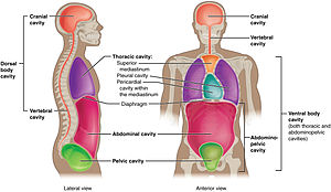
The body consists of a number of different cavities, separated areas which house different organ systems. The brain and central nervous system reside in an area protected from the rest of the body by the blood brain barrier. The lungs sit in the pleural cavity. The intestines, liver, and spleen sit in the abdominal cavity
Height, weight, shape and other body proportions vary individually and with age and sex. Body shape is influenced by the distribution of muscle and fat tissue.[27]
Physiology[edit]
Human physiology is the study of how the human body functions. This includes the mechanical, physical, bioelectrical, and biochemical functions of humans in good health, from organs to the cells of which they are composed. The human body consists of many interacting systems of organs. These interact to maintain homeostasis, keeping the body in a stable state with safe levels of substances such as sugar and oxygen in the blood.[28]
Each system contributes to homeostasis, of itself, other systems, and the entire body. Some combined systems are referred to by joint names. For example, the nervous system and the endocrine system operate together as the neuroendocrine system. The nervous system receives information from the body, and transmits this to the brain via nerve impulses and neurotransmitters. At the same time, the endocrine system releases hormones, such as to help regulate blood pressure and volume. Together, these systems regulate the internal environment of the body, maintaining blood flow, posture, energy supply, temperature, and acid balance (pH).[28]
Development[edit]
Development of the human body is the process of growth to maturity. The process begins with fertilisation, where an egg released from the ovary of a female is penetrated by sperm. The egg then lodges in the uterus, where an embryo and later fetus develop until birth. Growth and development occur after birth, and include both physical and psychological development, influenced by genetic, hormonal, environmental and other factors. Development and growth continue throughout life, through childhood, adolescence, and through adulthood to senility, and are referred to as the process of ageing.
Society and culture[edit]
Professional study[edit]

Health professionals learn about the human body from illustrations, models, and demonstrations. Medical and dental students in addition gain practical experience, for example by dissection of cadavers. Human anatomy, physiology, and biochemistry are basic medical sciences, generally taught to medical students in their first year at medical school.[29][30][31]
Depiction[edit]

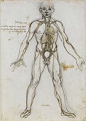
Anatomy has served the visual arts since Ancient Greek times, when the 5th century BC sculptor Polykleitos wrote his Canon on the ideal proportions of the male nude.[32] In the Italian Renaissance, artists from Piero della Francesca (c. 1415–1492) onwards, including Leonardo da Vinci (1452–1519) and his collaborator Luca Pacioli (c. 1447–1517), learnt and wrote about the rules of art, including visual perspective and the proportions of the human body.[33]
History of anatomy[edit]

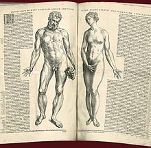
In Ancient Greece, the Hippocratic Corpus described the anatomy of the skeleton and muscles.[34] The 2nd century physician Galen of Pergamum compiled classical knowledge of anatomy into a text that was used throughout the Middle Ages.[35] In the Renaissance, Andreas Vesalius (1514–1564) pioneered the modern study of human anatomy by dissection, writing the influential book De humani corporis fabrica.[36][37] Anatomy advanced further with the invention of the microscope and the study of the cellular structure of tissues and organs.[38] Modern anatomy uses techniques such as magnetic resonance imaging, computed tomography, fluoroscopy and ultrasound imaging to study the body in unprecedented detail.[39]
History of physiology[edit]
The study of human physiology began with Hippocrates in Ancient Greece, around 420 BC,[40] and with Aristotle (384–322 BC) who applied critical thinking and emphasis on the relationship between structure and function. Galen (c. 126–199) was the first to use experiments to probe the body's functions.[41][42] The term physiology was introduced by the French physician Jean Fernel (1497–1558).[43] In the 17th century, William Harvey (1578–1657) described the circulatory system, pioneering the combination of close observation with careful experiment.[44] In the 19th century, physiological knowledge began to accumulate at a rapid rate with the cell theory of Matthias Schleiden and Theodor Schwann in 1838, that organisms are made up of cells.[43] Claude Bernard (1813–1878) created the concept of the milieu interieur (internal environment), which Walter Cannon (1871–1945) later said was regulated to a steady state in homeostasis.[40] In the 20th century, the physiologists Knut Schmidt-Nielsen and George Bartholomew extended their studies to comparative physiology and ecophysiology.[45] Most recently, evolutionary physiology has become a distinct subdiscipline.[46]
See also[edit]
- Medicine – The science and practice of the diagnosis, treatment, and prevention of physical and mental illnesses
- Glossary of medicine
- Body image
- Cell physiology
- Comparative physiology
- Comparative anatomy
- Development of the human body
References[edit]
- ^ "Chemical Composition of the Human Body". About education. Retrieved 2 September 2016.
- ^ "Fluid Physiology". Anaesthesiamcq. Retrieved 2 September 2016.
- ^ Ganong's 2016, p. 5.
- ^ "The Cells in Your Body". Science Netlinks. Retrieved 2 September 2016.
- ^ a b c Ron Sender; Shai Fuchs; Ron Milo (2016). "Revised estimates for the number of human and bacteria cells in the body". PLOS Biology. 14 (8): e1002533. bioRxiv 036103. doi:10.1371/journal.pbio.1002533. PMC 4991899. PMID 27541692.
- ^ Bianconi, E; Piovesan, A; Facchin, F; Beraudi, A; Casadei, R; Frabetti, F; Vitale, L; Pelleri, MC; Tassani, S; Piva, F; Perez-Amodio, S; Strippoli, P; Canaider, S (5 July 2013). "An estimation of the number of cells in the human body". Annals of Human Biology. 40 (6): 463–471. doi:10.3109/03014460.2013.807878. PMID 23829164.
- ^ David N., Fredricks (2001). "Microbial Ecology of Human Skin in Health and Disease". Journal of Investigative Dermatology Symposium Proceedings. 6: 167–169. doi:10.1046/j.0022-202x.2001.00039.x. Retrieved 7 February 2017.
- ^ Ganong's 2016, p. 16.
- ^ "Gene Expression | Learn Science at Scitable". www.nature.com. Retrieved 2017-07-29.
- ^ "tissue – definition of tissue in English". Oxford Dictionaries | English. Retrieved 2016-09-17.
- ^ a b Gray's Anatomy 2008, p. 27.
- ^ "organ | Definition, meaning & more | Collins Dictionary". www.collinsdictionary.com. Retrieved 2016-09-17.
- ^ "Cardiovascular System". U.S. National Cancer Institute. Archived from the original on 2 February 2007. Retrieved 2008-09-16.
- ^ Human Biology and Health. Upper Saddle River, NJ: Pearson Prentice Hall. 1993. ISBN 0-13-981176-1.
- ^ "The Cardiovascular System". SUNY Downstate Medical Center. 8 March 2008.
- ^ "Your Digestive System and How It Works". NIH. Retrieved 4 September 2016.
- ^ "Hormonal (endocrine) system". Victoria State Government. Retrieved 4 September 2016.
- ^ Zimmerman, Kim Ann. "Immune System: Diseases, Disorders & Function". LiveScience. Retrieved 4 September 2016.
- ^ Integumentary+System at the US National Library of Medicine Medical Subject Headings (MeSH)
- ^ Marieb, Elaine; Hoehn, Katja (2007). Human Anatomy & Physiology (7th ed.). Pearson Benjamin Cummings. p. 142.
- ^ Zimmerman, Kim Anne. "Lymphatic System: Facts, Functions & Diseases". LiveScience. Retrieved 4 September 2016.
- ^ Moore, Keith L.; Dalley, Arthur F.; Agur Anne M.R. (2010). Moore's Clinically Oriented Anatomy. Phildadelphia: Lippincott Williams & Wilkins. pp. 2–3. ISBN 978-1-60547-652-0.
- ^ "Nervous System". Columbia Encyclopedia (6th ed.). Columbia University Press. 2001. ISBN 978-0-7876-5015-5.
- ^ "Introduction to the Reproductive System". Epidemiology and End Results (SEER) Program. Archived from the original on 2 January 2007.
- ^ Maton, Anthea; Hopkins, Jean Susan; Johnson, Charles William; McLaughlin, Maryanna Quon; Warner, David; LaHart Wright, Jill (2010). Human Biology and Health. Prentice Hall. pp. 108–118. ISBN 0-13-423435-9.
- ^ Zimmerman, Kim Ann. "Urinary System: Facts, Functions & Diseases". LiveScience. Retrieved 4 September 2016.
- ^ Gray, Henry (1918). "Anatomy of the Human Body". Bartleby. Retrieved 4 September 2016.
- ^ a b "What is Physiology?". Understanding Life. Retrieved 4 September 2016.
- ^ "Introduction page, "Anatomy of the Human Body". Henry Gray. 20th edition. 1918". Retrieved 27 March 2007.
- ^ Publisher's page for Gray's Anatomy. 39th edition (UK). 2004. ISBN 0-443-07168-3. Archived from the original on 20 February 2007. Retrieved 27 March 2007.
- ^ "Publisher's page for Gray's Anatomy. 39th edition (US). 2004. ISBN 0-443-07168-3". Archived from the original on 9 February 2007. Retrieved 27 March 2007.
- ^ Stewart, Andrew (November 1978). "Polykleitos of Argos," One Hundred Greek Sculptors: Their Careers and Extant Works". Journal of Hellenic Studies. 98: 122–131. doi:10.2307/630196. JSTOR 630196.
- ^ "Leonardo". Dartmouth College. Retrieved 2 September 2016.
- ^ Gillispie, Charles Coulston (1972). Dictionary of Scientific Biography. VI. New York: Charles Scribner's Sons. pp. 419–427.
- ^ Hutton, Vivien. "Galen of Pergamum". Encyclopædia Britannica 2006 Ultimate Reference Suite DVD.
- ^ "Vesalius's De Humanis Corporis Fabrica". Archive.nlm.nih.gov. Retrieved 29 August 2010.
- ^ "Andreas Vesalius (1514–1567)". Ingentaconnect. 1 May 1999. Retrieved 29 August 2010.
- ^ "Microscopic anatomy". Encyclopædia Britannica. Retrieved 14 October 2013.
- ^ "Anatomical Imaging". McGraw Hill Higher Education. 1998. Retrieved 25 June 2013.
- ^ a b "Physiology – History of physiology, Branches of physiology". www.Scienceclarified.com. Retrieved 2 September 2016.
- ^ Fell, C.; Griffith Pearson, F. (November 2007). "Thoracic Surgery Clinics: Historical Perspectives of Thoracic Anatomy". Thorac Surg Clin. 17 (4): 443–448, v. doi:10.1016/j.thorsurg.2006.12.001.
- ^ "Galen". Discoveriesinmedicine.com. Retrieved 29 August 2010.
- ^ a b Newman, Tim. "Introduction to Physiology: History And Scope". Medicine News Today. Retrieved 2 September 2016.
- ^ Zimmer, Carl (2004). "Soul Made Flesh: The Discovery of the Brain – and How It Changed the World". J Clin Invest. 114 (5): 604–604. doi:10.1172/JCI22882. PMC 514597.
- ^ Feder, Martin E. (1987). New directions in ecological physiology. New York: Cambridge Univ. Press. ISBN 978-0-521-34938-3.
- ^ Garland, Jr, Theodore; Carter, P. A. (1994). "Evolutionary physiology" (PDF). Annual Review of Physiology. 56 (1): 579–621. doi:10.1146/annurev.ph.56.030194.003051. PMID 8010752.
Books[edit]
- Ganong's Review of Medical Physiology. 2016. ISBN 978-0-07-182510-8.
- Gray's anatomy: the anatomical basis of clinical practice. Editor-in-chief, Susan Standring (40th ed.). London: Churchill Livingstone. 2008. ISBN 978-0-8089-2371-8.
External links[edit]
| Wikisource has original works on the topic: Human Anatomy |
| Wikimedia Commons has media related to Human body. |
| Look up body in Wiktionary, the free dictionary. |
| Wikibooks has a book on the topic of: Human Physiology |
- The Book of Humans (from the late 18th and early 19th centuries)
- Inner Body
- Anatomia 1522–1867: Anatomical Plates from the Thomas Fisher Rare Book Library
.svg/80px-Diagram_of_the_human_heart_(cropped).svg.png)


.jpg/220px-Baby-256x385_1839565_640(1).jpg)
